GUIDEYE station
For everyone working with medical images
Research presentations, papers, teaching, and personal reflection.
Having DICOM pooled on your PC will be a great asset in your clinical career.

Not approved by the Ministry of Health, Labor and Welfare. Not for use in diagnostic or therapeutic purposes.

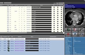
Database Features
This product is equipped with a database function and can read all DICOM data, including XP, XA, CT, MRI, NM (scintigraphy), US, IVUS, and OCT, and organize and save it by series.
By pooling DICOM data on a PC in advance, you can convert any data into still images or videos when creating slides for presentations at academic conferences. This dramatically improves the efficiency of presentation preparation. Furthermore, past data can be easily referenced, making it useful in discussions.
Cine viewer
With a single shortcut key, you can hide all patient and facility information. This can be used for conference presentations and other presentations.
Additionally, notes recorded in the database are displayed at the top of the screen, allowing you to grasp the characteristics and important points of an image at a glance.
In addition, XA, US, IVUS, and OCT images can be played back smoothly on the cine viewer, allowing for more intuitive image confirmation and analysis.


Generic viewer
Various images such as CT, MRI, and X-rays can be displayed.
In particular, for CT images, volume rendering is automatically generated.
The displayed 3D image can also be edited as is, making it extremely useful for visual analysis and document creation.
CT MPR
For CT images, MPR (multiplanar reconstruction) analysis and slab MIP (maximum intensity projection) creation are possible.
By utilizing MPR analysis, various measurements can be performed flexibly.
In addition, you can intuitively adjust the window width/window level (WW/WL) and smoothly perform view operations such as panning, zooming in and out.

Generic viewer

Various images such as CT, MRI, and X-rays can be displayed.
In particular, for CT images, volume rendering is automatically generated.
The displayed 3D image can also be edited as is, making it extremely useful for visual analysis and document creation.
Database Features

Searchable
You can add and manage notes for each series or even study, and you can also search by keyword.
The imported DICOM data is automatically stored in a unique database, so there is no need to store the original data separately.
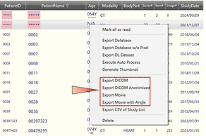
Output
In addition to being saved in DICOM format (which can also be anonymized), data can also be output as video (MP4) or still images.
You can use the data flexibly according to your needs.
Edit, save
Analyzed data is saved as a series and can be easily recalled at any time.
Furthermore, analysis data can be shared and transferred to other GUDEYE Station users.
This analysis data does not contain pixel information, so it does not put a strain on storage capacity and allows for efficient operation.
The imported DICOM data is automatically saved in a unique database, so there is no need to store the original data separately.
Cine viewer
Ideal for slide TM
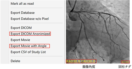
It is possible to embed angle information in the output angiographic images. This is extremely useful when making presentations at academic conferences or creating materials.
In addition, a unique hash value is automatically assigned to the output image, allowing you to quickly identify the original DICOM data (the display of the hash can also be set to hidden).
This means you don't have to remember which data the video or still image you pasted into your slide comes from.
Dual-screen synchronized playback
It is possible to embed angle information in the output angiographic images. This is extremely useful when making presentations at academic conferences or creating materials.
In addition, a unique hash value is automatically assigned to the output image, allowing you to quickly identify the original DICOM data (the display of the hash can also be set to hidden).
This means you don't have to remember which data the video or still image you pasted into your slide comes from.

It can be announced as is.
It is possible to embed angle information in the output angiographic images. This is extremely useful when making presentations at academic conferences or creating materials.
In addition, a unique hash value is automatically assigned to the output image, allowing you to quickly identify the original DICOM data (the display of the hash can also be set to hidden).
This means you don't have to remember which data the video or still image you pasted into your slide comes from.
Grasp the three-dimensional image
It allows for automatic angle-synchronized display of analyzed CT and XA images, making it effective for post-operative reviews.
About Purchase
[Please read before applying.]
This program is for research and educational use only. It is not for use in diagnostic or therapeutic purposes.
We assume no responsibility for any use other than for research and education.
Please check the operating environment requirements on this website before applying.